Manhattan Macular Hole Treatment
A macular hole is a breakdown in the macula, which is in the center of the retina. A macular hole can cause your sharp, central vision to become blurry and distorted. Macular holes are related to aging and usually occur in patients over the age of 50. A macular hole is different from a macular pucker, and different from age-related macular degeneration. Your eye doctor will know the difference between these conditions.
How Does a Macular Hole Occur?
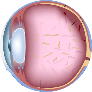
A macular hole occurs as the vitreous pulls away from the retina as we age. The vitreous is the gel-like substance that fills the interior of the eye. As we age, the vitreous slowly shrinks and pulls away from the retinal surface. Natural fluids fill the space that is left, and in most cases, there are no adverse effects. Some patients may experience an increase in floaters, which are small specks that appear to float around in the patients field of vision.
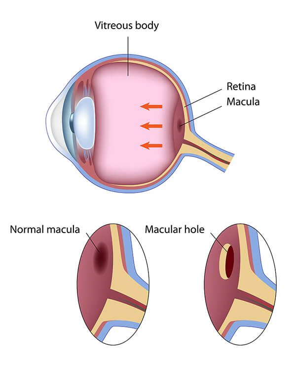
If the vitreous is firmly attached to the retina, the pulling away can tear the retina and create a macular hole. Also, once the vitreous has pulled away from the surface of the retina, some of the vitreous fibers can remain of the retinal surface and can contract. This causes tension on the retina and can lead to a macular hole. In both cases, the fluid that replaces the shrunken vitreous can seep through the hole onto the macula, blurring and distorting central vision.
What are the Symptoms of Macular Hole?
Macular holes often begin gradually. In the early stages, patients may notice a slight distortion or blurriness in their straight-ahead vision. Straight lines or objects can begin to look bent or wavy. Reading and performing other routine tasks can become difficult with the affected eye.
How do you Treat Macular Hole?
Some macular holes can heal themselves, but a vitrectomy is often necessary in many cases to improve vision. During this procedure, the vitreous gel is removed to prevent it from pulling on the retina and replaced with a bubble containing a mixture of gas and air. This bubble acts as an internal bandage that hold the edge of the macular hole in place until it heals.
Following surgery, patients must remain in a face-down position, normally for a day or two, but sometimes for two to three weeks. This position allows the bubble to press against the macular hole, and be gradually reabsorbed by the eye, sealing the hole. Maintaining a face-down position is crucial to the success of this surgery. The most common risk of vitrectomy is an increase in cataract development. Full recovery can often take up to three months.
If you are experiencing the symptoms of a macular hole, make sure to see your doctor or one of our doctors at Angioletti Retina. The longer you have a macular hole, the less chance you have to regain normal vision. Give Angioletti Retina a call and schedule your appointment today!